Bedridden Patient Height and Weight Estimation
- Last formula update:
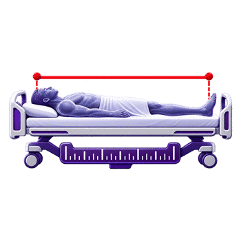
Bedridden Patient Height Calculator
Height estimation
Weight estimation
Results
Decimal & Rounding Policy
- Convert selected units before calculating; keep full precision throughout the formulas.
- Show estimated height and weight with up to two decimal places; omit unnecessary trailing zeros.
- Show reverse-solved editable measurements with up to 12 significant digits.
- Forearm lookup accepts 0.5 cm steps and returns the chart’s listed height without interpolation.
Valid range
- Semi-span: 50–115 cm.
- Arm length: 20–60 cm.
- Recumbent height: 100–230 cm.
- Knee height: 30–70 cm.
- Forearm length: 18.5–32 cm in 0.5 cm steps.
- Demispan: 50–115 cm.
- Age: 18–120 years overall; Cereda: 30–55 years; age-adjusted knee-height method: 60–90 years.
- Forearm chart age band: under 65 or 65 and older.
- Arm circumference: 15–60 cm.
- Calf circumference: 15–80 cm.
- Abdominal circumference: 40–180 cm.
- Subscapular skinfold: 2–80 mm.
- Editable or calculated height: 100–230 cm; weight: 20–300 kg.
- These are calculator entry bounds, not validated ranges for every prediction equation.

Brinelle Lumquell
Reviewers:
Cavarine Pexmont
Yeldric Vossbourne
Check our editorial policy
September 27, 2026
1.0.0
Initial calculator and formula release.
Our engineers are here to help you get it right.
How can a bedridden patient height calculator help when standing measurement is impossible?
Bedridden Patient Height Calculator estimates adult stature from reachable body segments and weight from selected circumferences. Check for a recent reliable direct measurement first. Otherwise, choose a method whose landmarks and study group fit the person. Enter each observed value with its unit. The tool applies the chosen equation or forearm chart.
- Knee, forearm, semi-span, demispan, and recumbent measures support different height routes.
- Arm, calf, and abdominal measures support weight routes; some need skinfold.
- The forearm chart uses half-centimeter rows and age bands.
- Reverse solving infers one missing measurement when other inputs are known.
- Record the method, input, unit, date, and estimated status.
The Bedridden Patient Height Calculator does not directly measure height or weight. Methods may disagree because their inputs, study groups, and coefficients differ. Check landmarks, position, units, edema, and contractures before interpreting a surprising answer. Use professional judgment, and replace an estimate with a reliable direct measure when feasible. A displayed decimal is calculation detail, not a promise of individual precision. The chart provides discrete values; it cannot fill gaps between rows. Keep the chosen branch visible when the result enters another calculation or handoff.
Assumptions used in this calculator
- The calculator is intended for adults within each method’s stated age range.
- Segment lengths and circumferences are converted to centimeters before estimation.
- Subscapular skinfold is converted to millimeters before weight estimation.
- Age is converted to years before age-dependent calculations.
- Anatomical landmarks are measured consistently by a trained observer.
- Knee height follows the selected method’s measurement protocol.
- Sex coding follows each equation’s original coefficient definition.
- White and Black branches retain the original study categories.
- The ulna lookup uses only listed half-centimeter measurements.
- An exact chart match is required for reverse ulna lookup.
- Reverse solving assumes exactly one missing numerical measurement.
- Edema or contractures may reduce the reliability of estimates.
- Results require clinical interpretation before use in care decisions.
Results are rounded for display.
Internal calculations use full precision.
Formulas Used in Bedridden Patient Height :
Input unit conversion
vbase = ventered × f
Mitchell and Lipschitz height
H = 2 × S
WHO semi-span height
H = 43 + 1.46 × S
Rabito height, first equation
H1 = 58.694 − 2.974 × G − 0.0736 × A + 0.4958 × L + 1.132 × S
Rabito height, second equation
H2 = 63.525 − 3.237 × G − 0.06904 × A + 1.293 × S
Gray recumbent height
H = R
Chumlea height, White men
H = 71.85 + 1.88 × K
Chumlea height, Black men
H = 73.42 + 1.79 × K
Chumlea height, White women
H = 70.25 + 1.87 × K − 0.06 × A
Chumlea height, Black women
H = 68.1 + 1.86 × K − 0.06 × A
Cereda height
H = 60.76 + 2.16 × K − 0.06 × A + 2.76 × Q
Age-adjusted knee height, men
H = 64.19 − 0.04 × A + 2.02 × K
Age-adjusted knee height, women
H = 84.88 − 0.24 × A + 1.83 × K
Forearm height lookup
H = chart value at (U, sex, age band)
Use the listed 0.5 cm row and selected chart column; do not interpolate.
Demispan height, men
H = 57.8 + 1.4 × D
Demispan height, women
H = 60.1 + 1.35 × D
Ross weight, White men
W = 1.19 × K + 3.21 × M − 86.82
Ross weight, Black men
W = 1.09 × K + 3.14 × M − 83.72
Ross weight, White women
W = 1.01 × K + 2.81 × M − 66.04
Ross weight, Black women
W = 1.24 × K + 2.81 × M − 82.48
Chumlea weight, men
W = 0.98 × C + 1.16 × K + 1.73 × M + 0.37 × T − 81.69
Chumlea weight, women
W = 1.27 × C + 0.87 × K + 0.98 × M + 0.4 × T − 62.35
Rabito weight, first equation
W1 = 0.503 × M + 0.5634 × B + 1.318 × C + 0.0339 × T − 43.156
Rabito weight, second equation
W2 = 0.4808 × M + 0.5646 × B + 1.316 × C − 42.245
Rabito weight, third equation
W3 = 0.5759 × M + 0.5263 × B + 1.2452 × C − 4.8689 × G − 32.9241
Selected output unit and display
vdisplay = formatup to 2 decimals(vbase ÷ f)
No intermediate formula result is rounded.
Reverse solve for one missing linear input
X = Y − b − ∑(ai × xi)aX
The sum includes all known terms; calculate in base units and validate the inferred input.
Reverse forearm chart lookup
U = exact chart row matching (H, sex, age band)
Reverse lookup succeeds only when the height appears in the selected column.
Variables & Definitions
View a complete list of all variables used in this calculator, including definitions and units
Variables Used in Bedridden Patient Height and Weight Estimation
| Symbol | Meaning | Unit or coding |
|---|---|---|
| H, H1, H2 | Estimated height; first and second Rabito height estimates | cm |
| W, W1, W2, W3 | Estimated weight; three Rabito weight estimates | kg |
| S | Semi-span | cm |
| L | Arm length | cm |
| R | Recumbent height | cm |
| K | Knee height | cm |
| U | Forearm or ulna length | cm |
| D | Demispan | cm |
| A | Age | years |
| M | Arm circumference | cm |
| C | Calf circumference | cm |
| B | Abdominal circumference | cm |
| T | Subscapular skinfold thickness | mm |
| G | Sex code in Rabito equations | male = 1; female = 2 |
| Q | Sex code in the Cereda equation | male = 1; female = 0 |
| Sex, age band | Selected categories for the forearm chart | male/female; under 65/65+ |
| v, f | Measured or estimated value; selected unit factor to its base unit | base units: cm, mm, kg, years |
| X, Y, b | Unknown measurement; edited result; equation intercept in reverse solving | units follow the selected equation |
| aX, ai, xi | Unknown's coefficient; known coefficients; known measurements | coefficients use the equation's base units |
Unit Conversion Table
Bedridden Patient Height and Weight Unit Conversion Table
| Unit Group | Unit Name | Symbol | Equivalent in Base Unit | Used For |
|---|---|---|---|---|
| Length | Centimeter | cm | 1 cm = 1 cm | Body segments and circumferences |
| Length | Inch | in | 1 in = 2.54 cm | Body segments and circumferences |
| Length | Millimeter | mm | 1 mm = 0.1 cm | Body segments and circumferences |
| Length | Meter | m | 1 m = 100 cm | Body segments and circumferences |
| Stature | Centimeter | cm | 1 cm = 1 cm | Recumbent and estimated height |
| Stature | Meter | m | 1 m = 100 cm | Recumbent and estimated height |
| Stature | Inch | in | 1 in = 2.54 cm | Recumbent and estimated height |
| Stature | Foot | ft | 1 ft = 30.48 cm | Recumbent and estimated height |
| Stature | Millimeter | mm | 1 mm = 0.1 cm | Recumbent and estimated height |
| Skinfold | Millimeter | mm | 1 mm = 1 mm | Subscapular skinfold thickness |
| Skinfold | Centimeter | cm | 1 cm = 10 mm | Subscapular skinfold thickness |
| Skinfold | Inch | in | 1 in = 25.4 mm | Subscapular skinfold thickness |
| Mass | Kilogram | kg | 1 kg = 1 kg | Estimated or edited weight |
| Mass | Pound | lb | 1 lb = 0.45359237 kg | Estimated or edited weight |
| Mass | Stone | st | 1 st = 6.35029318 kg | Estimated or edited weight |
| Mass | Gram | g | 1 g = 0.001 kg | Estimated or edited weight |
| Age | Year | yr | 1 yr = 1 year | Age-based equations |
| Age | Month | mo | 1 mo = 1/12 year | Age-based equations |
Example Calculation
Male patient · age 70 years · semi-span 80 cm · arm length 35 cm
H1 = 58.694 − 2.974(1) − 0.0736(70) + 0.4958(35) + 1.132(80) = 158.481 cm
H2 = 63.525 − 3.237(1) − 0.06904(70) + 1.293(80) = 158.8952 cm
Displayed heights: 158.48 cm and 158.9 cm
The first Rabito equation uses arm length as well as age and semi-span.
The second uses age and semi-span without arm length.
Both are estimates expressed in centimeters.
Only the displayed values are rounded; the equations retain full precision.
White male branch · entered weight 68.98 kg · knee height 50 cm · arm circumference missing
W = 1.19K + 3.21M − 86.82
M = (68.98 + 86.82 − 1.19 × 50) ÷ 3.21 = 96.30 ÷ 3.21
Inferred arm circumference: 30 cm
The entered weight and knee height leave one unknown measurement.
Rearranging the selected Ross equation gives arm circumference.
The inferred value lies within the calculator's 15–60 cm arm range.
A reverse-solved measurement is mathematical inference, not an observed measurement.
Results are rounded for display.
Internal calculations use full precision.
Calculations Disclaimer
When the Chart Has No Reliable Standing Height
A patient cannot stand today, and the chart lists an old height. A bedridden patient height calculator can help organize the next measurement. It estimates stature from body segments that staff can reach safely. The bedridden patient height calculator can also estimate weight from selected circumferences. Both outputs depend on the method and the quality of each input.
Start with a practical question: which measurement can you obtain without strain? A knee, forearm, or arm may be accessible while standing remains impossible. A recent, trustworthy direct height may be more useful than a new estimate. Record the source of any value. An estimate should never silently replace a measured entry in the chart.
Check for a Recent Measured Height First
An admission note may show a height, but its origin is often unclear. Ask whether the value was measured, reported, copied, or estimated. A recent direct measure deserves attention when it is reliable and relevant. A decades-old value may describe an earlier body shape or posture. The date matters, especially for older adults.
If direct measurement is possible, follow the local procedure. Otherwise, document why an alternative was needed. Keep the estimate beside its method and date. This helps the next reader avoid treating a prediction as an observation. It also makes later comparison possible when standing or recumbent measurement becomes feasible.
Ask who recorded the older number and what device was used. An old field may have been copied between visits. That does not make it useless, but its provenance is uncertain. When a fresh direct value cannot be obtained, keep this distinction visible in the record.
Choose a Reachable Body Segment
A fixed knee makes knee height difficult to obtain. An accessible forearm may then be more useful. A painful shoulder can make a reach-based measurement less reliable. The safest choice depends on the person’s position, mobility, and available equipment.
The calculator offers separate routes, not a universal ranking of methods. Some use one length. Others combine age, sex coding, and several body measurements. Select a route only after checking its required inputs. Do not force a limb into position to satisfy an equation. A comfortable, repeatable measure gives the calculation a better starting point.
One staff member may reach the forearm easily. Another may obtain a reliable knee measure with a suitable instrument. The best practical route changes with equipment and patient comfort. State why a route was chosen when the distinction might matter later.
How to Measure the Body Segments
A one-centimeter tape error can change a result before any rounding occurs. Place the tape at the correct landmarks and record the chosen unit. Repeat a difficult measure if the patient can tolerate it. Consistency matters more than adding digits to an uncertain input.
Keep the patient supported during the process. Explain each movement and protect privacy. If a position cannot be achieved safely, use another method. The calculator converts supported units internally, but it cannot correct a misplaced tape. Clear landmark notes help another observer reproduce the measurement later.
- Confirm the landmark and body position.
- Measure and record the raw value with its unit.
- Select the matching method and study branch.
- Check the output against the patient’s context.
- Document the estimate and its source.
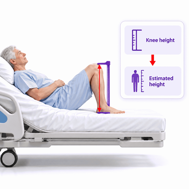
Knee Height with a Stable Right Angle
A knee held at an uneven angle can produce a misleading length. The usual knee-height procedure positions the knee and ankle at right angles. Measure the distance using the method’s intended landmarks and instrument. Avoid compressing soft tissue or guessing the heel endpoint.
Several equations use knee height, but their other inputs differ. One branch uses age and sex. Another uses historical population categories. A third combines knee height with age in a middle-aged cohort. Read the selected method before entering a number. A valid numerical input alone does not establish that the equation fits the person.
Forearm Length Between Elbow and Wrist
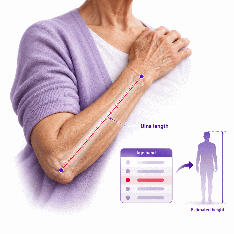
Wrist jewelry or a hidden elbow point can shift an ulna measurement. Find the olecranon at the elbow. Then locate the prominent ulnar styloid at the wrist. Measure between those landmarks in centimeters. The forearm chart uses half-centimeter rows, so record the appropriate row carefully.
Choose the chart column for the selected sex and age band. In this calculator, age 65 belongs to the older band. The result is a lookup value, not a continuous regression. A value between rows is not calculated by drawing a line through the chart. Repeat the measurement if a row choice feels uncertain.
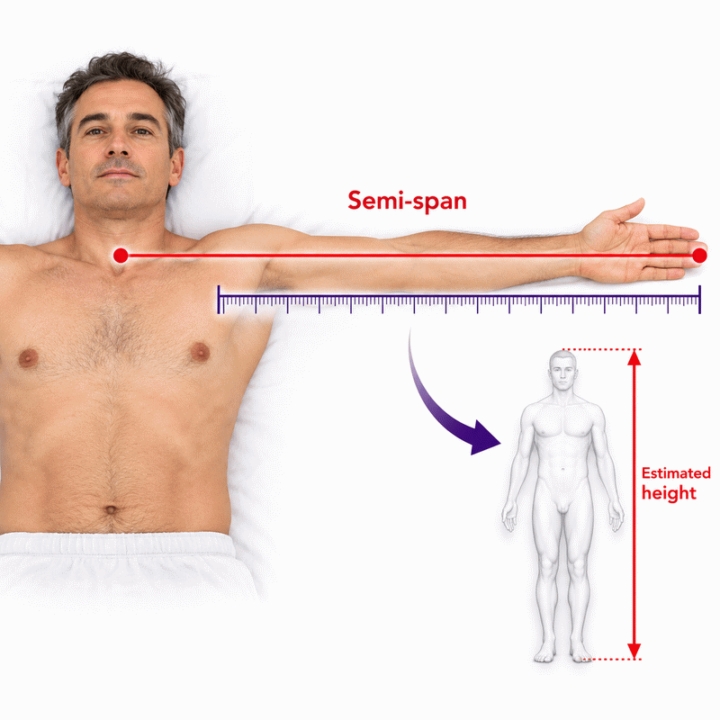
Semi-Span and Demispan from the Sternal Notch
An arm may not extend level with the shoulder. That changes a reach-based length. For the relevant methods, measure from the sternal notch toward the middle fingertip. Keep the arm in the intended position when it is safe to do so. Record any limitation that affects reach.
The calculator has both semi-span and demispan options because the selected equations differ. One simple model doubles semi-span. Another applies a coefficient and intercept. Sex-specific demispan equations use their own coefficients. Equal input lengths can therefore yield different predicted heights. These differences reflect different models, not a change in the physical tape measure.
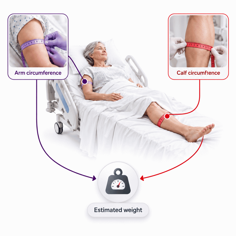
Arm Length and Circumferences for Weight Methods
A sleeve can hide an arm landmark or press against the tape. Arm length and arm circumference are separate measurements. Arm length contributes to one height equation. Mid-upper arm circumference contributes to several weight equations. Measure around the marked mid-arm point for circumference.
Calf and abdominal circumferences have different roles. Take each at the prescribed site without pulling the tape tight. A skinfold is a thickness, measured in millimeters. It is not interchangeable with a circumference in centimeters. Fluid shifts, dressings, and body position can affect the readings. Note those conditions before relying on a weight prediction.
Which Height Method Fits Your Available Inputs?
A form offers many study names, but only one may match today’s measurements. Choose by accessible anatomy and the study’s context. The simpler route may need one segment. A multi-input route may use age, sex coding, and arm length. More inputs do not automatically create a more accurate individual estimate.
Begin with the measurement you trust most. Then check whether the equation’s age span and group definitions apply. Keep the chosen method visible when recording the answer. If two routes are available, their difference can prompt a review of technique. It should not be averaged without a clinical reason.
For a patient with a comfortable elbow and a fixed knee, a forearm route may be feasible. For another patient, direct recumbent length may be clearer. Neither choice follows from the formula alone. The patient’s position, available equipment, and purpose of the estimate all influence the decision.
Direct Recumbent Length and Its Limits
A person can lie flat but cannot stand. Crown-to-heel measurement may then be possible. The recumbent option displays the entered length as height. It does not run a regression or adjust for mattress conditions.
The apparent simplicity can hide measurement error. Check the head and heel landmarks, body alignment, and position of the tape. Contractures and spinal curvature may make a straight length difficult. A flexible tape across bedding may not represent the same path as a rigid height measure. Label the result according to how it was obtained. Do not assume a displayed decimal makes the observation exact.
Semi-Span Models: Doubling and Regression
A single semi-span length can feed two different routes. The Mitchell and Lipschitz option doubles it. The WHO-labeled option applies a linear regression. The calculator uses centimeters for both inputs and outputs.
H = 2 × S
H = 43 + 1.46 × S
For a semi-span of 80 cm, these models give 160 cm and 159.8 cm. Neither figure is a direct measure of standing height. The agreement here is a property of this input value. It does not prove either method’s accuracy for a particular patient. Check that the notch and fingertip were found consistently before comparing results.
Knee Height Equations by Age and Study Group
A knee measure can be clear while the matching equation is not. The calculator separates several knee-height methods. Some branches use sex and historical White or Black study groups. The age-adjusted knee-height route applies to the studied 60–90-year span. Cereda’s route uses age, knee height, and sex coding for ages 30–55.
These coefficients came from particular populations. A group label describes a study branch, not a biological rule for every person. Review the clinical context before choosing a branch. When the patient’s situation differs substantially, document that limitation. A precise calculation does not remove model uncertainty.
Forearm Chart and Age Band
The tape reads 26.5 cm, but the chart column still needs selection. Pick the age band and sex column before reading the row. The tool displays that row’s listed height. It does not fit a new equation to the chart.
The chart is especially convenient when the lower limbs are hard to position. Convenience does not make the answer a measured stature. Population differences, landmark error, and unusual body proportions can change agreement. Record “ulna-based estimated height” with the input and chart band. If the forearm cannot be measured at its intended endpoints, use another available route.
A chart value can look more certain than a regression because it has a printed row. Yet every row still represents an estimated relationship. The chart cannot observe spinal curvature, current posture, or every individual’s proportions. Treat its listed centimeter value as a documented estimate.
Demispan and Arm Length Options
A patient can extend an arm, but knee positioning remains painful. Demispan provides a sex-specific height option. The Rabito routes use semi-span and age, with arm length added to the first equation. The second Rabito height can appear before arm length is entered because it needs fewer inputs.
This partial result can help staff avoid guessing a missing arm length. A later arm measure enables the other result. Do not interpret two displayed values as repeated measurements of the same thing. They are outputs from distinct equations. Note which one was used downstream. Recheck an implausible arm length before treating the difference as a clinical finding.
Estimating Weight When a Bed Scale Is Unavailable
A bed scale is unavailable, but a weight entry is requested. Tape-based equations can provide an estimate from reachable measurements. The calculator keeps weight estimation separate from height estimation. It cannot weigh the patient or detect rapid fluid change.
Choose a method whose inputs can be measured consistently. Some need knee height and arm circumference. Others add calf circumference, abdominal circumference, or subscapular skinfold. The result uses kilograms internally. Check the selected output unit before copying it. A change in edema, dressings, or tape placement can change the estimate even if usual tissue mass has not changed.
Knee and Arm Circumference Route
Only the knee and upper arm can be measured today. The Ross route uses those two values with the selected historical branch. Its coefficients differ across the original study categories. The arithmetic is linear, so a larger measured circumference raises its predicted weight when other inputs stay fixed.
For one White male branch, knee height 50 cm and arm circumference 30 cm yield 68.98 kg. This is a worked calculation, not an observed patient. The result should be recorded as an estimate from that branch. If either tape value is doubtful, remeasure before using the output.
W = 1.19 × 50 + 3.21 × 30 − 86.82 = 68.98 kg
Calf, Arm, Knee, and Skinfold Route
A calf circumference is available, but a scale still is not. The Chumlea weight method combines calf, knee, and arm measures with a subscapular skinfold. Men and women use different coefficient sets. The skinfold belongs in millimeters; the lengths and circumferences belong in centimeters.
Look for swelling or a dressing around the calf. These can alter the circumference without mirroring ordinary body mass. A skinfold reading also depends on technique. Repeat a doubtful measurement and record any impediment. A small decimal on the screen should not hide uncertainty in the original tape or caliper reading.
Abdominal Circumference and Three Weight Results
Skinfold thickness cannot be obtained, but arm, calf, and abdominal circumferences can. The Rabito method can still show its second and third weight results. Its first result appears when skinfold is entered. The three equations use different coefficients and should remain distinct.
The third equation subtracts its sex-coded term and a constant of 32.9241. It does not create an additional positive-constant result. The model was developed and validated in particular hospitalized groups. Circumference changes from fluid, equipment, or position can affect transfer to another setting. Choose and record the specific equation rather than treating the three outputs as a vote.
When two results are available early, the missing skinfold is still missing. Do not manufacture it to make the first result appear. The partial calculation is a useful feature because it shows only what the current evidence supports.
Reading Results Without Mistaking an Estimate for a Measurement
Two outputs look precise, yet neither is guaranteed to equal standing height. The screen’s decimal places describe display formatting. They do not measure the model’s individual error. Keep the raw segment, method, and unit with the result.
A value can be mathematically correct for an equation and still be unsuitable for care. That distinction matters when results feed nutrition screening or other calculations. A clinician should assess whether the chosen model fits the patient and task. Replace an estimate with a reliable direct measure when one becomes available. Preserve the earlier estimate as a documented historical calculation.
- Measured segment: an observed tape or caliper value.
- Predicted stature or weight: a model output.
- Clinical use: a separate decision that considers uncertainty.
This three-step distinction prevents a common handoff error. A downstream spreadsheet may show a height but hide its origin. Carry the estimate label forward whenever that height supports another calculation.
Why Methods Can Disagree
Knee height suggests one stature, while demispan suggests another. Each model weights a different body segment and may come from a different study group. Age, posture, body proportions, and measurement technique also matter. A disagreement is a reason to investigate, not to average automatically.
First, verify units and landmarks. Next, check the chosen age and study branch. Then ask whether edema, contracture, or limited reach affected an input. If the gap remains, keep both values labeled and seek local clinical guidance. A difference between methods does not identify which one is correct for the individual.
Documentation and Handoff
A colleague receives “height 170 cm” with no context. That number could be measured, recalled, or predicted. Record the method name, each relevant input, its unit, and the calculation date. Mark the result as estimated.
A brief note might say that ulna length was measured to the nearest 0.5 cm. It should include the selected chart band. A knee-based note should state the chosen equation branch. This small amount of detail makes the result auditable. It also helps the next shift decide whether to repeat a measure. Never erase uncertainty by copying only the final figure.
Using the Online Tool in a Care Workflow
A measurement is ready, but the wrong study is selected. Change the method before entering values. The fields update to show what that route needs. Enter only measurements that were actually taken. Results update as valid inputs become available.
AxiCalculator provides a quick way to compare the calculator’s supported methods. The tool can convert supported units and export a record. Review that record before sharing it. For any decision with medical consequences, follow local procedures and professional judgment. The calculator assists the workflow; it cannot replace direct assessment.
Use the result screen as a check, not a shortcut past measurement notes. The share link records selected values and units for review. A receiving colleague should still verify the entered data. The downloadable record can help with communication when the team keeps its normal documentation process.
Review Inputs, Units, and Reverse Solving
A result is known, but one predictor is missing. The calculator can solve one unknown in a linear equation when its other terms are known. Enter the known result in its editable field after clearing the missing measurement. The inferred input is an algebraic value, not a tape reading.
For the ulna chart, reverse lookup needs an exact height in the selected column. An arbitrary height cannot be converted into a new chart row. Unit changes should preserve the underlying physical value. Check the displayed unit and the input limit after solving. If the inferred measure looks implausible, do not present it as observed.
What to Do When a Value Looks Implausible
An implausible result often begins with a simple input mistake. Check whether inches were entered as centimeters. Check whether skinfold thickness was entered in millimeters. Confirm the selected sex code, chart band, and study branch. Revisit the anatomical landmarks before changing an equation.
A second worked calculation shows the unit path. Enter a semi-span of 80 cm in the doubling method. The result is 160 cm. Enter the same physical length as 31.4961 inches, and the result remains about 160 cm. The displayed decimals may differ because the inch value was shortened.
Keep the distinction clear: measured length, converted input, predicted result, and clinical interpretation are separate steps. If a value still conflicts with the patient’s record, pause its use. A qualified clinician can select a better measure or document the uncertainty. Explore the AxiCalculator tool with a measurement you can verify.
Frequently Asked Questions
What if the patient's left forearm cannot be accessed?
Can repeat visits use this tool to track a patient's height change?
How should I interpret an estimate in severe spinal curvature?
Why does an export include more than one result?
Can a reverse-solved circumference satisfy a missing clinical measurement?
What if a direct height and a segment estimate disagree?
How should a historical population branch be used for someone outside that group?
Our engineers are here to help you get it right.